Compact Bone Diagram Lacunae / Compact Bone Spongy Bone And Other Bone Components Human Anatomy And Physiology Lab Bsb 141 / Compact bone is the denser, stronger of the two types of bone tissue (figure 6).
byAdmin-
0
Compact Bone Diagram Lacunae / Compact Bone Spongy Bone And Other Bone Components Human Anatomy And Physiology Lab Bsb 141 / Compact bone is the denser, stronger of the two types of bone tissue (figure 6).. Due to its structure, it is referred to as cortical bone. Usually found in long bones of the body, it consists of units called osteons, each of which is. The two tissues serve different purposes in bones, with. (on textbook page diagrams note only highlighted labels) compact bone. What is the difference between compact bone and spongy bone?
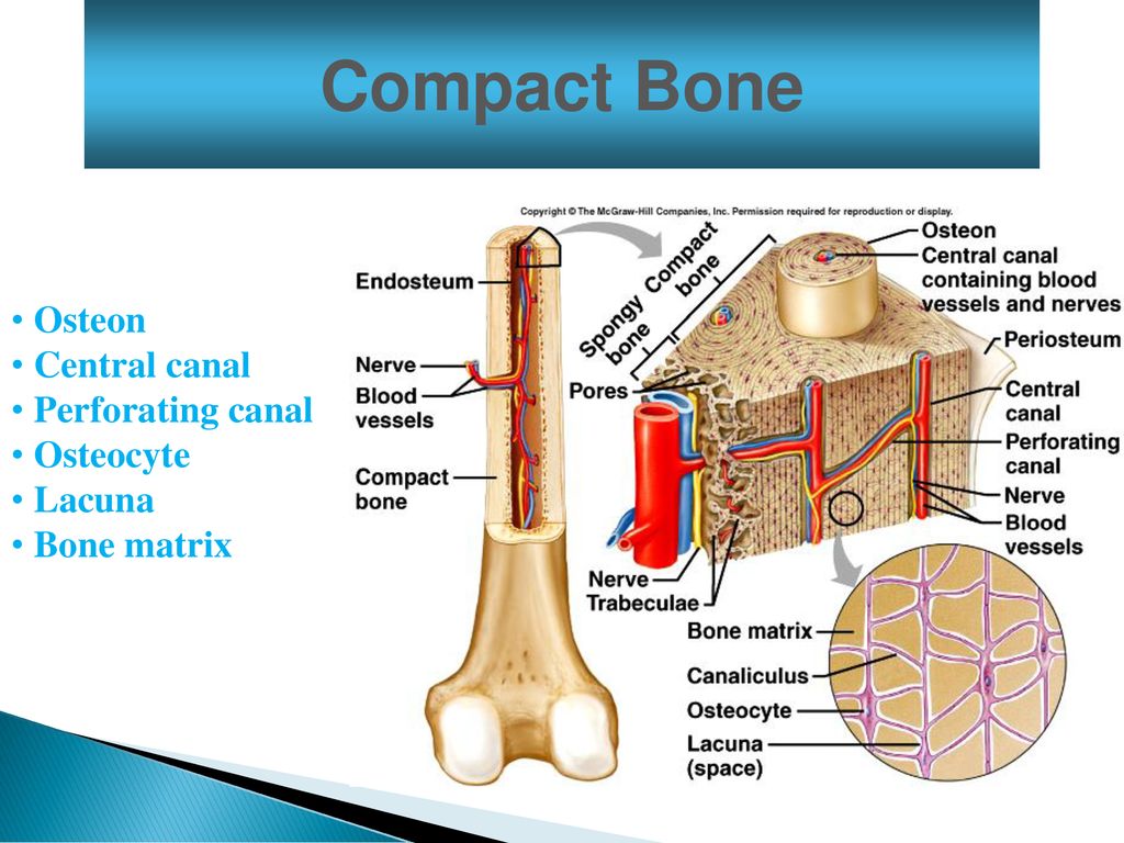
Like compact bone, spongy bone, also known as cancellous bone, contains osteocytes housed in lacunae, but they are not arranged in concentric circles. What is the difference between compact bone and spongy bone? List the 4 types of bones based on their shapes and list 2 examples of each type. Concentric lamellae interstitial lamellae central canal lacuna osteocyte canaliculus. Some, mostly older, compact bone is remodelled to form these haversian systems (or osteons).the osteocytes sit in their lacunae in concentric rings around a central haversian canal (which runs longitudinally).the osteocytes are arranged in concentric rings of bone matrix called lamellae (little plates), and their processes run in interconnecting canaliculi.
Bone Structure Ppt Download from slideplayer.com Compact bone is the denser, stronger of the two types of osseous tissue (figure 6.3.6). Between the rings of matrix, the bone cells (osteocytes) are located in spaces called lacunae. Concentric lamellae interstitial lamellae central canal lacuna osteocyte canaliculus. In histology a lacuna is a small space containing an osteocyte in bone or chondrocyte in cartilage. (the dark purple structures in the diagram above are the lacunae.) the osteocytes or mature bone cells are located in the lacunae. About press copyright contact us creators advertise developers terms privacy policy & safety how youtube works test new features press copyright contact us creators. An osteocyte within a lacuna. (on textbook page diagrams note only highlighted labels) compact bone.
Compact bone makes up 80.
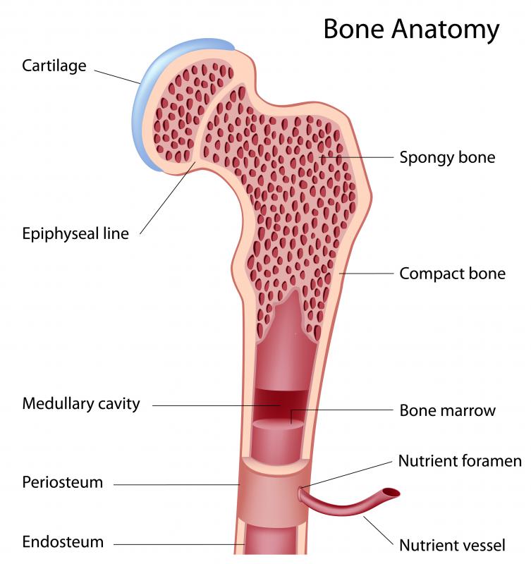
Link the lacunae _____ e. Between the rings of matrix, the bone cells (osteocytes) are located in spaces called lacunae. Human bone generally comprises osseous tissue, an outer coating called a periosteum, and bone marrow.the two main structural components typically include spongy bone on the interior, with an outer layer of compact bone. The osteon consists of a central canal called the osteonic (haversian) canal, which is surrounded by concentric rings (lamellae) of matrix. Answer there are two types of bone tissue : There are pores and spaces even in compact bone. An osteocyte within a lacuna. Compact bone consists of closely packed osteons or haversian systems. Noun plural lacunae luh kyoo nee ləˈkyu ni lacunas. Start studying compact bone labeling. Compact bone is the denser, stronger of the two types of bone tissue (figure 6). Osteon model lacunae canaliculi osteocyte. About press copyright contact us creators advertise developers terms privacy policy & safety how youtube works test new features press copyright contact us creators.
Due to its function, compact bone is also referred to as strong bone; Andrew kirmayer a diagram of the anatomy of a bone, showing the compact bone. There are pores and spaces even in compact bone. Compact bone forms the surface of all bones. Do you want to learn the details of the histology of compact bone with labelled diagram and authentic slide images?
What Is The Function Of Compact Bone With Pictures from images.infobloom.com Lacunae are small cavities or chambers located between one lamella and the next. Learn vocabulary, terms, and more with flashcards, games, and other study tools. Compact bone accounts for 80% of the bones in the human body. Compact bone is the denser, stronger of the two types of osseous tissue (figure 6.3.6). Osteocytes receive nutrients and eliminate wastes through blood vessels in the compact bone. Link the lacunae _____ e. An enlarged view of an osteon showing the ostecytes within lacunae and the concentric lamellae. Answer there are two types of bone tissue :
In long bones, as you move from the outer cortical compact bone to the inner medullary cavity, the bone transitions to spongy bone.
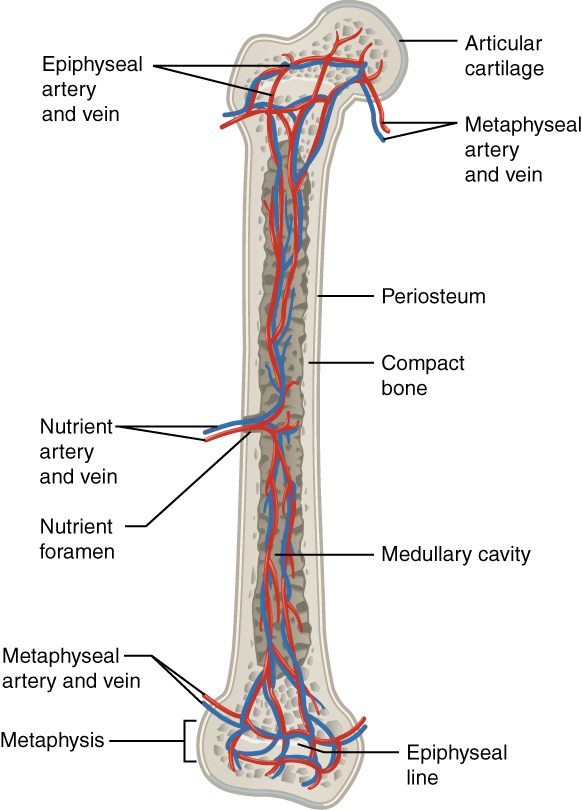
Lacunae are small cavities or chambers located between one lamella and the next. Compact bone makes up 80. The diaphysis and the epiphysis. Good, here in this part, i am going to describe the structure of compact bone. An air space in the cellular tissue of plants. Answer there are two types of bone tissue : An osteocyte within a lacuna. A structural unit of compact bone consisting central haversian canal. Compact bone is the denser, stronger of the two types of osseous tissue (figure 6.3.6). Usually found in long bones of the body, it consists of units called osteons, each of which is. Compact bone is the denser, stronger of the two types of bone tissue (figure 6). It can be found under the periosteum and in the diaphyses of long bones, where it provides support and protection. About press copyright contact us creators advertise developers terms privacy policy & safety how youtube works test new features press copyright contact us creators.
A structural unit of compact bone consisting central haversian canal. The remainder is cancellous bone, which has a spongelike appearance with numerous large spaces and is found in the. An osteocyte within a lacuna. A thin layer of compact bone also covers the epiphyses of long bones. Andrew kirmayer a diagram of the anatomy of a bone, showing the compact bone.
What Is The Structure And Function Of The Compact Bone Socratic from opentextbc.ca Learn vocabulary, terms, and more with flashcards, games, and other study tools. It can be found under the periosteum and in the diaphyses of long bones, where it provides support and protection. An air space in the cellular tissue of plants. A structural unit of compact bone consisting central haversian canal. Andrew kirmayer a diagram of the anatomy of a bone, showing the compact bone. Compact bone histology slide structure with diagram. The two tissues serve different purposes in bones, with. Like compact bone, spongy bone, also known as cancellous bone, contains osteocytes housed in lacunae, but they are not arranged in concentric circles.
Good, here in this part, i am going to describe the structure of compact bone.
Do you want to learn the details of the histology of compact bone with labelled diagram and authentic slide images? Osteocyte is an entrapped osteoblast in the matrix. Compact bone, also called cortical bone, dense bone in which the bony matrix is solidly filled with organic ground substance and inorganic salts, leaving only tiny spaces (lacunae) that contain the osteocytes, or bone cells.compact bone makes up 80 percent of the human skeleton; Like compact bone, spongy bone, also known as cancellous bone, contains osteocytes housed in lacunae, but they are not arranged in concentric circles. Usually found in long bones of the body, it consists of units called osteons, each of which is. In long bones, as you move from the outer cortical compact bone to the inner medullary cavity, the bone transitions to spongy bone. Human bone generally comprises osseous tissue, an outer coating called a periosteum, and bone marrow.the two main structural components typically include spongy bone on the interior, with an outer layer of compact bone. Due to its structure, it is referred to as cortical bone. A thin layer of compact bone also covers the epiphyses of long bones. The canaliculi give the osteon the appearance of having tiny cracks in the lamellae. Learn vocabulary, terms, and more with flashcards, games, and other study tools. Osteocytes, located in lacunae, are connected to one another by processes in canaliculi. Some, mostly older, compact bone is remodelled to form these haversian systems (or osteons).the osteocytes sit in their lacunae in concentric rings around a central haversian canal (which runs longitudinally).the osteocytes are arranged in concentric rings of bone matrix called lamellae (little plates), and their processes run in interconnecting canaliculi.
A diagram of the femur showing a cut through the compact bone into the medullary cavity compact bone diagram. Good, here in this part, i am going to describe the structure of compact bone.