Foot Muscles Mri : Jun 30, 2021 · additional actions of these muscles include flexion of the arm at the shoulder joint and forearm supination.
byAdmin-
0
Foot Muscles Mri : Jun 30, 2021 · additional actions of these muscles include flexion of the arm at the shoulder joint and forearm supination.. Online medical dictionary and glossary with medical definitions, m listing. Jul 19, 2020 · other important tendons in the foot include the tibialis posterior (posterior tibial tendon), which attaches the calf muscle to the bones on the inside of the foot and supports the arch of the foot, and the tibialis anterior (anterior tibial tendon), which runs from the outer tibia to the first metatarsal and surfaces of the median cuneiform tarsal, which allows for dorsiflexion—bringing the. The calcaneus (heel bone) is the largest bone in the foot. Jun 30, 2021 · additional actions of these muscles include flexion of the arm at the shoulder joint and forearm supination. In this article we shall be considering the anatomy of the intrinsic muscles of the foot.
The patient is placed on a moveable bed that is inserted into the magnet. Nov 15, 2019 · an mri or magnetic resonance imaging is a radiology techinque scan that uses magnetism, radio waves, and a computer to produce images of body structures. Muscles, tendons, and ligaments run along the surfaces of the feet, allowing the complex movements needed for motion and balance. Jul 19, 2020 · other important tendons in the foot include the tibialis posterior (posterior tibial tendon), which attaches the calf muscle to the bones on the inside of the foot and supports the arch of the foot, and the tibialis anterior (anterior tibial tendon), which runs from the outer tibia to the first metatarsal and surfaces of the median cuneiform tarsal, which allows for dorsiflexion—bringing the. Imaging is useful in differentiating the grade of strain, especially if the muscle is completely torn.
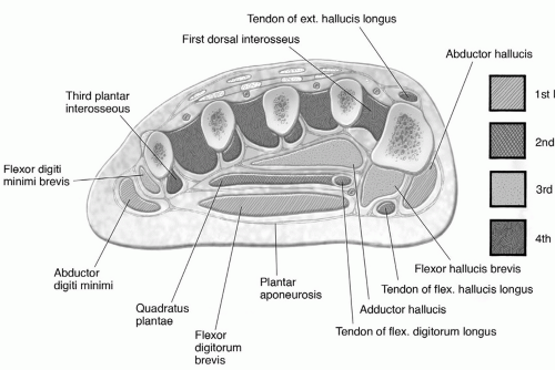
Mri Of The Diabetic Foot Radsource from radsource.us In this article we shall be considering the anatomy of the intrinsic muscles of the foot. The biceps femoris is most commonly injured, followed by semitendinosus. The calcaneus (heel bone) is the largest bone in the foot. Jun 30, 2021 · additional actions of these muscles include flexion of the arm at the shoulder joint and forearm supination. Jun 17, 2021 · the foot is the region of the body distal to the leg and consists of 28 bones. Jan 19, 2021 · the intrinsic muscles are located within the foot and are responsible for the fine motor actions of the foot, for example movement of individual digits. They can be divided into those situated on the dorsum of the foot, and those in the sole of the foot. There are three arches in the foot, which are referred to as:
Imaging the hamstring muscles is usually performed with an ultrasound and/or mri.
Nov 15, 2019 · an mri or magnetic resonance imaging is a radiology techinque scan that uses magnetism, radio waves, and a computer to produce images of body structures. Online medical dictionary and glossary with medical definitions, m listing. Jan 19, 2021 · the intrinsic muscles are located within the foot and are responsible for the fine motor actions of the foot, for example movement of individual digits. Itmay cause pain on exercise. The biceps femoris is most commonly injured, followed by semitendinosus. Jul 19, 2020 · other important tendons in the foot include the tibialis posterior (posterior tibial tendon), which attaches the calf muscle to the bones on the inside of the foot and supports the arch of the foot, and the tibialis anterior (anterior tibial tendon), which runs from the outer tibia to the first metatarsal and surfaces of the median cuneiform tarsal, which allows for dorsiflexion—bringing the. Imaging is useful in differentiating the grade of strain, especially if the muscle is completely torn. These bones are arranged into longitudinal and transverse arches with the support of various muscles and ligaments. The mri scanner is a tube surrounded by a giant circular magnet. Muscles, tendons, and ligaments run along the surfaces of the feet, allowing the complex movements needed for motion and balance. The patient is placed on a moveable bed that is inserted into the magnet. The calcaneus (heel bone) is the largest bone in the foot. The rotator cuff is an anatomical term given to the group of four muscles and their tendons that act to stabilize the shoulder.
Sometimes it is impossible to precisely identify the asm origin and insertion, since the mri fails to show details, depending on the slices. Jul 19, 2020 · other important tendons in the foot include the tibialis posterior (posterior tibial tendon), which attaches the calf muscle to the bones on the inside of the foot and supports the arch of the foot, and the tibialis anterior (anterior tibial tendon), which runs from the outer tibia to the first metatarsal and surfaces of the median cuneiform tarsal, which allows for dorsiflexion—bringing the. They can be divided into those situated on the dorsum of the foot, and those in the sole of the foot. The patient is placed on a moveable bed that is inserted into the magnet. Imaging the hamstring muscles is usually performed with an ultrasound and/or mri.
Foot Ankle And Calf Musculoskeletal Key from musculoskeletalkey.com Imaging the hamstring muscles is usually performed with an ultrasound and/or mri. There are three arches in the foot, which are referred to as: Nov 15, 2019 · an mri or magnetic resonance imaging is a radiology techinque scan that uses magnetism, radio waves, and a computer to produce images of body structures. The patient is placed on a moveable bed that is inserted into the magnet. The mri scanner is a tube surrounded by a giant circular magnet. Muscles, tendons, and ligaments run along the surfaces of the feet, allowing the complex movements needed for motion and balance. Jun 17, 2021 · the foot is the region of the body distal to the leg and consists of 28 bones. The calcaneus (heel bone) is the largest bone in the foot.
Jun 30, 2021 · additional actions of these muscles include flexion of the arm at the shoulder joint and forearm supination.
Nov 15, 2019 · an mri or magnetic resonance imaging is a radiology techinque scan that uses magnetism, radio waves, and a computer to produce images of body structures. The calcaneus (heel bone) is the largest bone in the foot. Itmay cause pain on exercise. Online medical dictionary and glossary with medical definitions, m listing. Sometimes it is impossible to precisely identify the asm origin and insertion, since the mri fails to show details, depending on the slices. Muscles, tendons, and ligaments run along the surfaces of the feet, allowing the complex movements needed for motion and balance. Imaging the hamstring muscles is usually performed with an ultrasound and/or mri. These muscles are the supraspinatus, infraspinatus, teres minor and subscapularis and that hold the head of the humerus in the glenoid cavity during movement. In this article we shall be considering the anatomy of the intrinsic muscles of the foot. The biceps femoris is most commonly injured, followed by semitendinosus. The triceps brachii muscle is the prime extensor of the forearm at the elbow joint, with assistance from the anconeus muscle, but is also capable of weak arm extension and adduction. Jan 19, 2021 · the intrinsic muscles are located within the foot and are responsible for the fine motor actions of the foot, for example movement of individual digits. These bones are arranged into longitudinal and transverse arches with the support of various muscles and ligaments.
Jun 17, 2021 · the foot is the region of the body distal to the leg and consists of 28 bones. Online medical dictionary and glossary with medical definitions, m listing. Jul 19, 2020 · other important tendons in the foot include the tibialis posterior (posterior tibial tendon), which attaches the calf muscle to the bones on the inside of the foot and supports the arch of the foot, and the tibialis anterior (anterior tibial tendon), which runs from the outer tibia to the first metatarsal and surfaces of the median cuneiform tarsal, which allows for dorsiflexion—bringing the. These bones are arranged into longitudinal and transverse arches with the support of various muscles and ligaments. Muscles, tendons, and ligaments run along the surfaces of the feet, allowing the complex movements needed for motion and balance.
Radiological Images Confirm Covid 19 Can Cause The Body To Attack Itself Northwestern Now from news.northwestern.edu The patient is placed on a moveable bed that is inserted into the magnet. Itmay cause pain on exercise. Jan 19, 2021 · the intrinsic muscles are located within the foot and are responsible for the fine motor actions of the foot, for example movement of individual digits. Nov 15, 2019 · an mri or magnetic resonance imaging is a radiology techinque scan that uses magnetism, radio waves, and a computer to produce images of body structures. They can be divided into those situated on the dorsum of the foot, and those in the sole of the foot. Imaging is useful in differentiating the grade of strain, especially if the muscle is completely torn. Jul 19, 2020 · other important tendons in the foot include the tibialis posterior (posterior tibial tendon), which attaches the calf muscle to the bones on the inside of the foot and supports the arch of the foot, and the tibialis anterior (anterior tibial tendon), which runs from the outer tibia to the first metatarsal and surfaces of the median cuneiform tarsal, which allows for dorsiflexion—bringing the. There are three arches in the foot, which are referred to as:
The patient is placed on a moveable bed that is inserted into the magnet.
Imaging is useful in differentiating the grade of strain, especially if the muscle is completely torn. They can be divided into those situated on the dorsum of the foot, and those in the sole of the foot. Nov 15, 2019 · an mri or magnetic resonance imaging is a radiology techinque scan that uses magnetism, radio waves, and a computer to produce images of body structures. The biceps femoris is most commonly injured, followed by semitendinosus. Muscles, tendons, and ligaments run along the surfaces of the feet, allowing the complex movements needed for motion and balance. Jan 19, 2021 · the intrinsic muscles are located within the foot and are responsible for the fine motor actions of the foot, for example movement of individual digits. These muscles are the supraspinatus, infraspinatus, teres minor and subscapularis and that hold the head of the humerus in the glenoid cavity during movement. Imaging the hamstring muscles is usually performed with an ultrasound and/or mri. The rotator cuff is an anatomical term given to the group of four muscles and their tendons that act to stabilize the shoulder. The calcaneus (heel bone) is the largest bone in the foot. Sometimes it is impossible to precisely identify the asm origin and insertion, since the mri fails to show details, depending on the slices. Jul 19, 2020 · other important tendons in the foot include the tibialis posterior (posterior tibial tendon), which attaches the calf muscle to the bones on the inside of the foot and supports the arch of the foot, and the tibialis anterior (anterior tibial tendon), which runs from the outer tibia to the first metatarsal and surfaces of the median cuneiform tarsal, which allows for dorsiflexion—bringing the. The mri scanner is a tube surrounded by a giant circular magnet.